Sectional Imaging
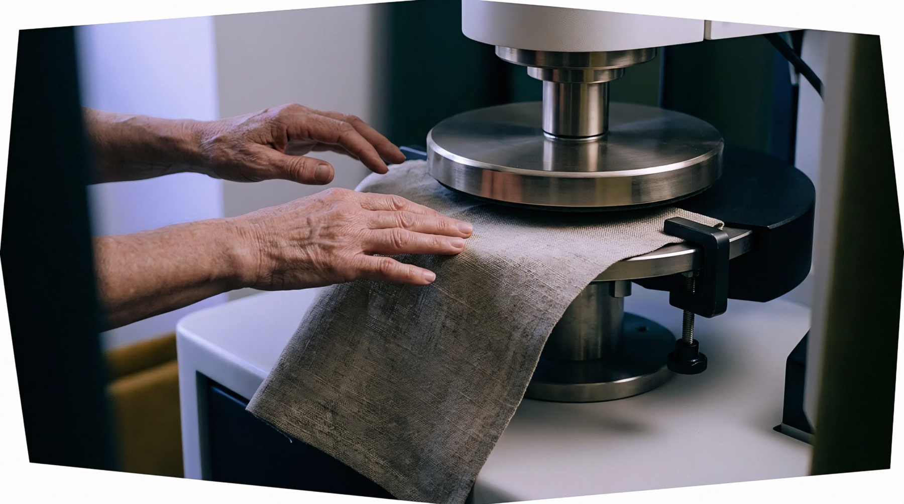
Microscopic analysis of sliced specimen planes isolates the internal morphology and arrangement of individual fibres within yarns or fabrics. Execution of transverse cross-section microscopy allows quality control technicians to inspect cell wall dimensions and resin penetration depth in linen textiles. Laboratory procedures mount embedded sample bundles in hardened resin matrix blocks before slicing perpendicular to the fibre axis.
Magnified images provide quantitative metrics for fibre wall area, lumen diameter and cross-sectional circularity.
Microtome Preparation
Technicians embed yarn or fabric samples in methacrylate or epoxy resins to preserve structural geometry during slicing operations. Precision rotary microtomes fitted with tungsten carbide blades cut ultra-thin sections measuring between two and five micrometers in thickness. Transferring slices onto glass slides allows high-power optical or scanning electron microscopes to resolve cellular details down to sub-micron scales.
Image analysis software calculates cross-sectional distribution, measuring total fibre area and counting individual bast ultimate cells within bundle structures. In blended yarns containing flax and polyester, section imaging quantifies component distribution across the yarn core and surface layers. Uniform dispersion of constituent fibres prevents uneven dye absorption and structural weak points in woven fabrics.
Inspection Limits
Preparation artifacts such as blade compression or resin shrinkage distort physical dimensions under high magnification. Sample preparation must strictly follow standardized embedding protocols to ensure valid quantitative dimensional measurements. Physical inspection remains limited to representative micro-samples selected from larger fabric production lots.